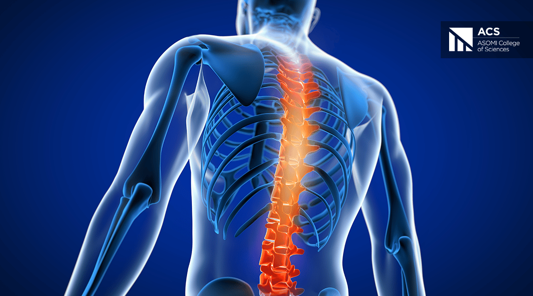
ASOMI College of Sciences is concerned with issues regarding health, including osteopathy. Therefore, ACS is honored to recommend the reading of the adaptation of a scientific paper by Mervyn Waldman on the principle of Tensegrity in osteopathy.
The article below is the third and the last part of this series of articles. Click here for the first and here for the second part. The part at hand includes also the biography.
Osteopathy – Tensegrity – Littlejohn
Any physical contact between two bodies is a stress transfer from one to another. In Osteopathy the stress transfer is seeking to be specifically utilizing appropriate vectors such as force, intensity, direction, duration, sequencing, velocity, and depth. The surface contact area is also crucial to ensure the appropriate level of strain. Whatever the effects, muscular, metabolic, neural, fascial, lymphatic, are all dependent on the appropriate level and delivery of the stress-guided transfer. The application of force on a targeted tissue translates into a change in spatial and textural properties of the matter that it impacts.
Physical therapies, dance, exercise, and various forms of movement can indeed influence cellular activities, including cell growth, differentiation, and potentially even immune cell responses that are critical to human health. Chemicals and genes are absolute of critical importance; however, they are governed by mechanical forces, as well as chemical cues. (Tozzi 2010, 2015)
The tensile and compression elements of tensegrity structures in the human body respond to the force of gravity vectors as they traverse through the body.
In the early decade of the 20th century, McConnel and Littlejohn began to formulate in increasing detail a proposed outline of the direction of the chief vectors and their potential influence on body mechanics and function.
Hohenschurz-Schmidt versus Littlejohn
Hohenschurz-Schmidt, in a recent article available on the web, contrasted the principles of tensegrity with those of Littlejohn’s study in body mechanics. I believe his analysis to be worthy of serious study and consideration.
He points out that, despite the apparent appearance of Littlejohn’s lines of force transmission through the body and their summation in polygons being a study in ‘statics’, in fact, throughout Littlejohn’s elaboration of the subject, it is ‘dynamics’ and the functional effects that dominate and underline its formulation.
Littlejohn emphasizes that it is the balance of spinal curve to curve relationships, vertebral body, and facet shapes, together with an analysis of transitional zones in the spinal column and the relation to the limbs, as an integrated motor unit, having to meet and adapt to gravitational forces, that determines human body mechanics.
To quote Hohenschultz-Schmidt:
“At a first glance, this spinal assessment has very little to do with a compression-tension system. Moreover, in Littlejohn’s writings, there are ‘pivots’, ‘levers’, and gravity-dependent force vectors. Advocates of a ‘bio-tensegrity’ make every attempt to ban these ‘traditional’ biomechanical terms from their analysis, stating that in a true tensegrity such levers could not exist, but that only non-linear force distribution along tensile trains could integrate focal compression points.
However, this vocabulary was, at the time of J.M.Littlejohn, the only one disposable. Littlejohn saw the T9 vertebra to be the keystone of a mainly compressive dorsal curvature; cervical and lumbar curves were deemed more ‘fluid’ in nature and bars of a keystone.
Nowadays, no serious biomechanical researcher would hold that the spine is a column of blocks, disregarding tension and lateral force transmission – such a notion has become the ‘bad guy or antagonist for many advocates of biotensegrity necessary to highlight the ‘uniqueness of their approach.
Besides that, even not J.M. Littlejohn considered the spine only as a compressive column; after a closer examination, Littlejohn was no less concerned with the soft-tissue influences on the spine than he was with joint shape and curve function.
For example, posterior and anterior cervical muscles were thought to influence the way C2 and C5 would move, (one being drawn posteriorly by the suboccipital musculature and the other tending to flex forward under the tension of the scalene and longus colli musculature).
The gravity lines constituting the polygon of forces have also been termed anterior and posterior ‘lines of articular tension’ (Campbell, undated), indicating that instead of a fixed compressive force, the body adapts along the lines to balance gravity. More importantly, the spine is not conceptualized as a column, instead, emphasis is made on particular points within a functional unit.
These points, either functional pivot or centers of gravity, could, alternatively, be conceptualized as areas of compression. They are isolated but functionally linked by the influence of tension. This tension can be conveyed through myofascial structures, thus, at least on a conceptual level its linkable to tensegrity”.

Bibliography
Hohenschurz-Schmidt, D., Littlejohn Tensegrity. http://osteopathstudent.weebly.com/littlejohn-tensegrity.html
Ingber, D., 1998. The Architecture of Life. Sci Am. 1998 Jan;278(1):48-57
Ingber, D. 2009. J Bodyw Mov Ther. 2008 Jul; 12(3): 198–200.
Langevin, H. M., 2008. Potential role of fascia in chronic musculoskeletal pain. In J. Audette, and A. Bailey (Eds). Integrative pain management: The science and practice of complementary and alternative medicine in pain management. (pp. 123-132). New Jersey, USA. Humana Press.
Levin, S. M., (1981) The icosahedron as a biologic support system, Proceedings, 34th Annual Conference on Engineering in Medicine and Biology, Huston
Levin, S.M., (2002) The tensegrity truss as a model for spine mechanics J Mechan Med Bio 2002; 2: 375-88L
Sharkey, John., (2015) The Fallacy of Biomechanics, http://www.johnsharkeyevents.com/blog/2015/3/11/biotensegrity-anatomy-for-the-21st-century
Stecco, C., Gagey, O., Belloni, A., 2007b. Anatomy of the deep fascia of the upper limb. Second part: study of innervations. Morphologie, 91, 38-43.
Stecco, A., Macchi, V., Stecco, C. Porzionato, A., Day, J.A., Delmas, V., De Caro, R., 2009. Anatomical study of myofascial continuity in the anterior region of the upper limb. Journal of Movement and Bodywork Therapies. 13(1). 53-62.
Standring, S., (2008). Gray’s Anatomy, The Anatomical Bases of Clinical Practice. (40th ed.). Edinburgh, United Kingdom: Elsevier Churchill Livingston.
Tozzi, Paolo., (2015). A unifying neuro-fasciagenic model of somatic dysfunction – underlying mechanisms and treatment – part 1 J. BodywMovTher. 2015 Apr; 19: 310-26
Tozzi, Paolo., et al (2010). Fascial release effects on patients with non-specific cervical or lumbar pain J. BodywMovTher. 2010 xx, 1-12
Van der Wal, J., 2009. The architecture of the connective tissue in the musculoskeletal system- an often overlooked functional parameters as to proprioception in the locomotor apparatus. International Journal of Therapeutic Massage and Bodywork. 2(4), 9-23.
Wood Jones, F., 1946. Buchanan’s Manual of Anatomy, 7th edn. London: Baill


